Diagnosis of Low Back Pain FAQs
Diagnosis of Low Back Pain FAQs
Written By DR CHOW Hung-Tsan
(Last update on: September 15th 2020)
How does one diagnose low back pain?
History:
Physical Examination:
X-rays:
Your doctor will usually arrange a series of X-rays. X-rays show the bones, but do not directly demonstrate soft tissues, however if disc degeneration is present, the X-rays will often show a narrowing of the intervertebral disc spaces between the vertebral bodies, which indicates the discs have become very thin or have collapsed. Bone spurs, called osteophytes, can also form around the edges of the vertebral bodies and around the edges of the facet joints. As the disc collapses and bone spurs form, the space available for the nerve roots can also start to shrink.
X-rays are often performed sitting or standing with the individual bending forwards (flexing) and leaning back (extending) to demonstrate abnormal motion, known as instability.
Magnetic Resonance Imaging (MRI):
MRI is a non-radiation technique for imaging the spine that uses a magnetic field. It can demonstrate soft tissue structures, such as the spinal cord, intervertebral discs (Fig. 1), etc as well as bone.
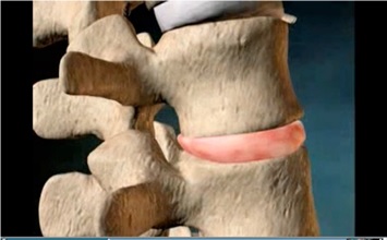 Fig. 1 Intervertebral disc |
MRI is the single most useful test available for diagnosing spinal disorders.
MRI images are used to examine intervertebral disc height, hydration and a disc’s ability to exchange nutrients with its vertebrae.
 |
| Fig. 2 The special weight bearing MRI scanner takes images with the individual’s positions: upright, tilting, & supine |
Computed Tomography (CT) Scan:
Bone Scan:
Myelogram:
Provocative Discography (PD):
What is the role of Discography? Is it enough to have MRI only?
In single level disc disease, the pain origin is usually correlated to the MRI findings, however there is a 30% chance that the MRI findings are not typically correlated with the symptom.
In multi-level disc disease, the situation is even more confusing. Each individual abnormal disc may present differently.
To establish the relationship between the abnormal findings in the MR images and the clinical presentation, we need discography to help us to make an accurate diagnosis of the pain origin. Usually your doctor can also treat the pain by therapeutic injection at the same time in performing the discography.
The most common discography procedure is Transforaminal Epidural Steroid Injection.
The advantages of discography are:
1. To correlate the relationship between the back pain symptom with the abnormal MRI findings. (To help your surgeon to select most appropriate choice of treatment for you) [2].
2. To exclude non-painful disc from multiple level disc degeneration (2-3 levels of lumbar disc degeneration are common, especially in older people).
3. Helping to decide which level needs surgery and how many levels need surgery [2].
4. Positive prediction value in predicting the prognosis of pain relief after surgery. In a study regarding treatment for low back pain, pre-operative positive discogram findings correlated with an 89% sustained clinical benefit from operative intervention, whereas negative discogram findings correlated with only 52% success post-operatively [2,3].
5. Pain injection therapy at the same time (for relieving the back pain – possibly preventing an operation).
6. Minimal risk and suitable to almost any patient, except those with contrast or local anaesthetic allergy and pregnant women.
How to do Lumbar Discography?
- You should not eat or drink for 2 hours before the procedure.
- You will be admitted to day surgery ward of the hospital.
- The procedure is done under local anaesthesia and you will be lying face down for 30 to 45 minutes (depending on how many levels and if additional procedures are required).
- The level of injection will be confirmed by X-ray screening.
- Your back will be covered with sterile drapes (Fig. 3).
- Your doctor will use lignocaine (a local anaesthetic) to numb your skin.
- Your doctor will pass a thin needle into the disc under X-ray guidance (Fig. 4).
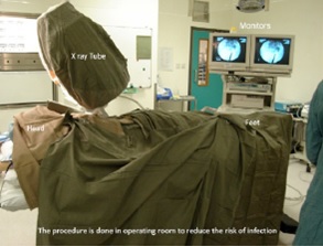 Fig. 3 Setting in the operating theatre for discogram. | |
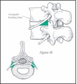 Fig. 4 The needle inserted into the disc space through a “safe zone”, labeled as green. | |
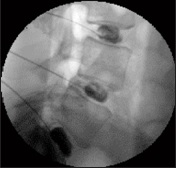 Fig. 5 Intra-operative images of discogram. |
The next step is the Relieving Test. Your doctor will inject lignocaine into the disc to see the pain relief response. We will record the change of the pain level one minute after the lignocaine injection. We might inject a normal disc for comparison if necessary. However, we try to avoid this to reduce the further discomfort and potential disc degeneration of a normal disc after puncture.
Additional pain-relieving procedures, such as epidural steroid injection, is usually performed during the discogram.
What should I do and expect after the procedure?
Twenty to thirty minutes after the procedure, you can move your back to try to provoke your usual pain. You will report your remaining pain, (if any) and also record the relief you experience during the next week. You may or may not obtain improvement in the first few hours after the injection, depending on if the disc injected is your main pain source. On occasion, you may feel numb, slightly weak or have an odd feeling in your leg for a few hours after the injection.
You may notice a slight increase in your pain lasting for several days as the numbing medication wears off before the cortisone is effective.
Ice will typically be more helpful than heat in the first 2-3 days after the injection.
You may begin to notice an improvement in your pain 2-5 days after the injection.
Occasionally, you may have some pain for longer than a week.
You may take your regular medications up to 2 weeks.
You may be referred for physical or manual therapy after the injection while the numbing medicine is effective and/or over the next several weeks while the cortisone is working.
Risks of Discography?
Risks of discography include discitis, neurologic and visceral injury, dye reactions, spinal headache and others. Spinal cord injury, vascular injury, pre-vertebral abscess, and subdural empyema have all been reported post-discography [4]. In 1995 it was recognized that the incidence of complications with discography were 0.15% per patient and 0.08% per disc [5].
The possibility that discography may cause persistent back pain since discography is generally performed in subjects with pre-existing back pain. Patients with significant emotional and psychological issues and chronic pain and those with disability claims account for more than 80% of those with persistent pain after experimental discography [6].
Selective Nerve Root Block (SNRB): This is injection test and/or therapeutic injection procedure for nerve root symptom (radiculopathy). This is done by injecting local anaesthetic and steroid on the nerve root under the X-ray guidance (Fig 6). The equipment, pre-procedure preparation and technique are identical to discography.
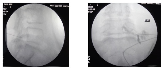 A B Fig. 6 Intra-operative Fluoroscopy of Selective Nerve Root Block Right L5 nerve root. A. Lateral view. B. Antero-posterior view |
Facet or Sacro-iliac Joint Injections:
Local anaesthetic injections into these joints are used to evaluate these joints as sources of low back pain. Judging by the effectiveness of the anaesthetic in relieving pain, a specific joint(s) producing back pain can be identified.Conclusion
Discography may prevent patients from undergoing unnecessary surgery.
In patients having an operation, post-operative success has been shown to improve with the use of preoperative discography, especially in cases of multi-level disc problems.
References
1. Palit M, Schofferman J, Goldthwaite N. Anterior Discectomy and Fusion for the Management of Neck Pain. Spine 1999; Vol.24 (21): 2224.
2. Simmons EH, Bhalla SK. Anterior Cervical Discectomy and Fusion. A clinical and biomechanical study with eight-year follow-up. J Bone Joint Surg (BR) 1969; 51: 225-37.
3. Colhoun E, McCall IW, Williams L, Pullicino VNC. Provocation discography as a guide to planning operations on the spine. J Bone Joint Surg (BR) 1988; 2:267-71.
4. Zeidman SM, Thompson K, Ducker TB. Complications of cervical discography: analysis of 4400 diagnostic disc injections. Neurosurgery. 1995; 37 (3): 414-7.
5. Guyer R, Ohnmeiss D. Lumbar discography. Position statement from the North American Spine: Society Diagnostic and Therapeutic Committee. Spine 1995; 20:2048-59.
6. Carragee EJ, Chen Y, Tanner C, Hayward C, Rossi M, Hagle C. Can discography cause long term back symptoms in previously asymptomatic subjects? Spine 2000; 25(14): 1803-1808.
Articles written/edited/reviewed by Doctors of Asia Medical Specialists ©2020 Asia Medical Specialists Limited. All rights reserved. |
