防治中风的新概念与最新发展
分享到
防治中风的新概念与最新发展
written by doctors of Asia Medical Specialists
(Last update on: Oct 28th 2020)
传统对「脑血管病变」的概念近年已被「脑中风」取替。与突发性心脏病一样,脑中风是属于突发性的心血管疾病,亦有相类似治疗概念。血管神经外科医生亦会先选择脑内血管介入疗法,而不是经静脉注射溶血剂。脑内血管介入疗法须于症状出现后的八小时内进行。这就是治疗脑中风最新的黄金八小时概念。
完整英文版本已经上载,中文版本即将更新。
摘要
“脑血管意外”的旧概念最近被“脑部攻击”的新概念所取代。就像心脏病发作一样,脑发作也是一种急性心血管疾病。心脏病发作和脑部发作的病因相似。心脏病发作的治疗概念也适用于脑部发作。对于
心脏病发作,心脏病专家更喜欢使用经皮冠状动脉介入治疗,即心脏PCI,而不是静脉注射重组组织纤溶酶原激活剂(例如iv rtPA)。症状发作后的12小时内必须进行心脏PCI。因此,IV rtPA是心脏病发作的第二线治疗,仅在PCI不可用时才提供。
类似的概念也适用于脑部发作。血管神经外科医生也更喜欢经皮脑介入术(Neuro-PCI),而不是静脉rtPA。症状发作后的8小时内必须执行Neuro-PCI。因此,这是用于脑部攻击的全新Golden-8小时概念。如今,Neuro-PCI是美国大多数综合卒中中心提供的一线标准治疗。 Golden-3-小时内的Iv rtPA被认为是第二线治疗,仅当Neuro-PCI不可用时才提供。
|
中风的新概念
脑血管意外的旧观念和对逐渐的神经系统恶化以及最终的残疾甚至死亡状态的命运的接受应该发生革命性的变化。新的术语“大脑发作”意味著中风不是偶然的。就像心脏病发作一样,中风是可以预防,治疗甚至治癒的,不会给患者带来任何神经系统缺陷。中风的治疗方法与心脏病发作类似。将来,患有急性缺血性中风的患者将更不会痛苦[1,2]。
什么是中风?
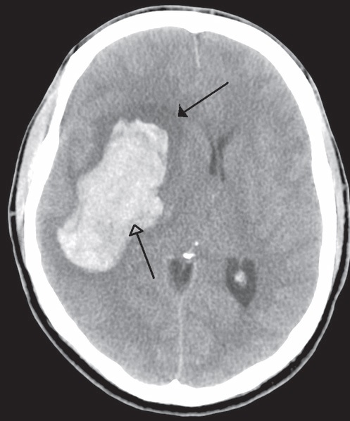
术语“中风”是指由于血管原因引起的神经功能障碍的突然发作。中风有两种类型:缺血性中风占西方世界所有中风的约87%(香港约占70%)。出血性中风解释了其余的13%(香港为30%)。但是出血性中风的死亡率高于缺血性实体。它占所有中风死亡的30%以上。Pathophysiology
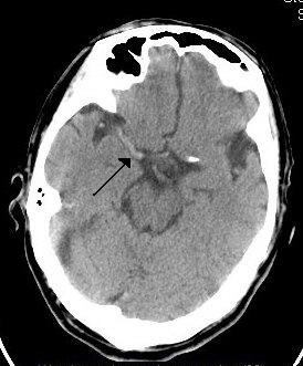
不论年龄,种族或性别,中风都会有机会发生在任何人身上。在缺血性中风期间,大脑中的血管被阻塞。阻塞会扰乱血液流动,阻止氧气输送到受影响的大脑区域。缺氧的脑组织部分变得“震惊”,不再正常工作,从而导致中风症状。
时间就是大脑
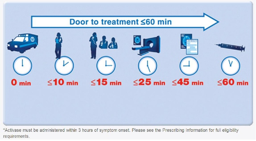
急性缺血性中风(AIS)是严重的医疗急症。快速介入至关重要。每分钟大血管中风得不到治疗,就会损失多达190万个神经元(每秒损失32,000个神经元,每小时损失1.2亿个神经元),失去140亿个突触,失去7.5英里的有髓纤维。中风得不到治疗的时间越长,永久性神经损伤的可能性就越大。如果延迟治疗,则“脑梗死”会导致关键脑细胞的额外死亡,从而降低治疗的潜在益处。短时间治疗对于增加良好恢复的机率绝对至关重要。大约三个小时后,脑细胞将遭受不可逆转的伤害,通常导致其死亡。这就是为什么仅在中风发作后三小时才进行rtPA溶栓治疗的原因。因此,时间确实是大脑[3].
中风对流行病学和社会经济影响
- 香港每年大约有18,000例中风病例。每年大约有795,000名美国人经历新的(610,000)或经常性(185,000)中风。
- 中风是美国第三大死亡原因,仅次于心脏病和癌症。据估计,每年约有60,000美国人死于中风。它导致全世界10%的死亡[4]。
- 由于人口老龄化,预计未来几十年中风的发病率将上升。它可能很快成为全世界最常见的死亡原因。[5].脑卒中的发病率从30岁开始呈指数增长,病因因年龄而异[6]。
- 中风是导致长期严重残疾的主要原因
- 中风是住院长期成人医疗保险报销的第一原因。现在,在美国,每年的中风总费用超过630亿美元。 2010年中风的直接和间接费用估计为737亿美元。
- 目前有超过300万人患有中风,其个人和经济损失是巨大的。
- 除身体和经济负担外,中风还给患者及其家人造成巨大的心理影响
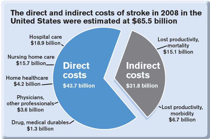 Fig. 2 Total stroke cost in US |
The New Concept of Stroke Management
The new concept emphasises screening of stroke risk, the control of risk factors and the prevention on brain attack; public education on stroke symptoms allowing rapid recognition and seeking of emergency help; and, most importantly, fast-track treatment of acute brain attack during the golden hours of the therapeutic window.
QT3
Question, Screening, Prevention, Time, Place, Person, 3D8P
(QSP-TPP-3D8P)
Screening for Stroke: Question, Screening, Prevention
- Question: Ask patients questions: their own estimation of life time stroke risk (reflecting their awareness of own health and family history)
- CLIS Screening: (Clinical screen, Laboratory screen, Image screen).
a. Clinical screen:
i. SDHHH History (personal and family): Stroke, Diabetes, Hyperlipidemia, Hypertension, Heart disease.
ii. BHCBP Examination: Body Mass Index, Heart status, Carotid artery for bruit, Blood pressure, Pulse for atrial fibrillation.
b. GOCO Laboratory screen: Glucose level, (Oil) Lipid profile, Clotting profile , (Organs) Liver and Renal function.
c. Image screen: Magnetic Resonance Imaging of brain, brain vessels and carotid and vertebral arteries. - Prevention of stroke or Risk factors control: (Up to 80% of strokes are preventable; you can prevent a stroke.
a. DEWCAD Lifestyle Modification: Diet (Calorie intake, Glucose, Fat, Salt consumption) [7], Exercise, Weight, Cigarettes 8 , Alcohol 9, Drug (Oral contraceptive pills, soft drugs [10])
b. POGAS Medical Therapy: Drug for Blood pressure control11, Cholesterolaemia [11], Glucose control for diabetes [12], Atrial Fibrillation (Anticoagulant)13, Stroke in the past (Antiplatelet)
c. Structural abnormalities with or without symptoms: Refer to vascular nerosurgeon for Prophylactic Neurosurgery Percutaneous Cerebral Intervention (Neuro-PCI)
i. Cervical arteries stenosis: i.e. carotid stent for carotid stenosis [14].
ii. Intracranial vascular anomaly: e.g. hypoplastic vessels in circle of Willis: only acknowledge and aware.
iii. Intracranial vascular stenosis: i.e. Intracranial vascular stent for Middle Cerebral Artery (MCA) stenosis [15].
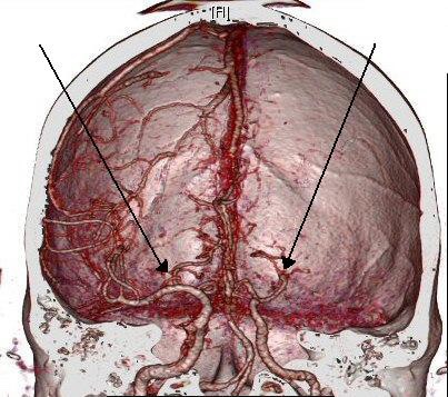
iv. Cerebral aneurysm or arteriovenous malformation: Endovascular embolisation therapy for reducing the risk of haemorrhagic stroke.
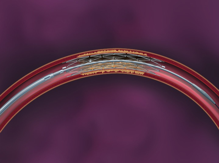 Fig. 3 The Wingspan system of intracranial vascular stent |
Management of Acute Stroke (TPP-3D8P)
Time--Place--Person--3 Golden hours Drug or PCI—8 Golden Hours only PCI (TPP-3D8P)
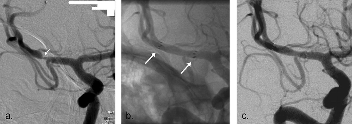 Fig. 4a The Pre-stenting angiogram (DSA) showing right MCA stenosis, b. stent-in-situ with stent marker (white arrow), c. the post-stenting DSA |
1. Time:
- Establish the exact time of symptoms onset or the time when patient was last seen to be normal. Recognition of stroke symptoms i.e. facial appearance, limbs weakness, speech problem,visual and sensory disturbance, gait and coordination, orientation and consciousness, etc.
- Promptly seek for emergency help during the Golden-3-Hours therapeutic window.
2. Place:
Seek immediate medical assessment and treatment in Hospital equipped with CT scanner, MRI scanner and Angiogram machine [16].
3. Person:
Seek help from vascular neurosurgeon who can provide comprehensive treatment and who can manage the complications associated with ischaemic stroke and its treatment.
4. 3-Golden-hours Drug or PCI:
- After clinical assessment of the contraindications for intravenous recombinant tissue plasminogen activator (iv rtPA or ‘Activase’), the fibrinolytic drug may be given as the first line treatment if the option of Neuro-PCI is not available.
- Most (>90%) of the acute ischaemic stroke patients will not benefit from iv rtPA due to: i. late presentation to emergency room (more than 2 hours after symptom onset), ii. fall into the exclusion criteria of iv rtPA, iii. delayed diagnosis in emergency rooms or clinical wards due to misinterpretation of CT scan.
- The benefit of iv rTPA should weighted against its associated high risks. Properly informed consent is vital [17, 18].
|
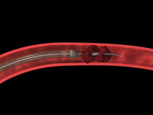
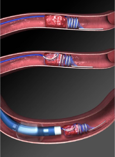
5. 8-Golden-Hours only PCI (Neurosurgical Percutaneous Cerebral Intervention)
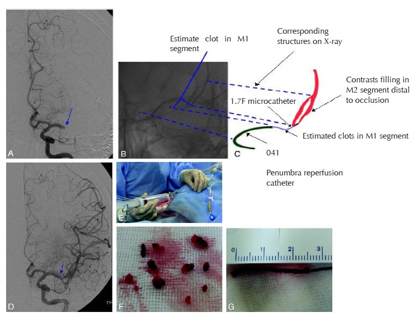
- Neuro-PCI achieves brain revascularisation by mechanical thrombectomy under fluoroscopic guidance of cerebral angiograms. A micro-catheter is passed up to the occluding thrombus in intracranial vessels. The thrombus or embolus is removed by the device using mechanical breakage and aspiration.
- Intra-arterial fibrinolysis i.e. ia rtPA injected at the site of thrombosis, improves outcomes in acute ischemic stroke [19] .
- Intra-vascular stent may be deployed if concomitant vascular stenosis exists.
- Within the Golden-3-hours, Neuro PCI can revascularise the brain cells and minimise the central infarction core. Patients may have full recovery without any neurological deficit.
- Within the Golden-8-Hours: From the therapeutic window of the 4th hour to the 7th hour, Neuro-PCI revascularisation can salvage brain cells in the peripheral ischaemic region called the Penumbra Zone and minimise the volume of brain infarction. Thus the overall morbidity and mortality of ischaemic stroke is reduced.
- Neuro-PCI is also indicated for patients who were unable to receive iv rtPA due to contraindications or for whom the drugs were ineffective [20, 21, 22, 23].
|
Salvage Neurosurgery
1. Surgery for bleeding complication of iv rtPA
Around 6% of iv rtPA treated patients develop symptomatic intracranial bleeding that cause more disastrous brain damage [24]. Salvage neurosurgery is difficult due to rtPA caused bleeding tendency. Though surgery may be lifesaving, patients may be severely disabled or even vegetative.
|

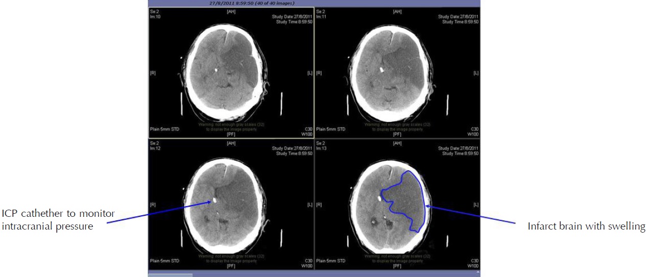
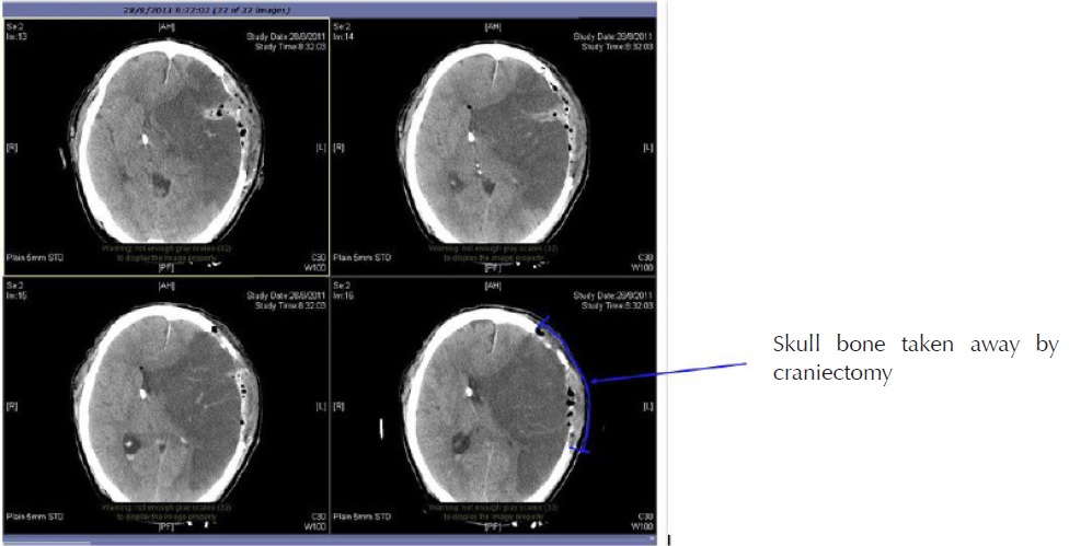
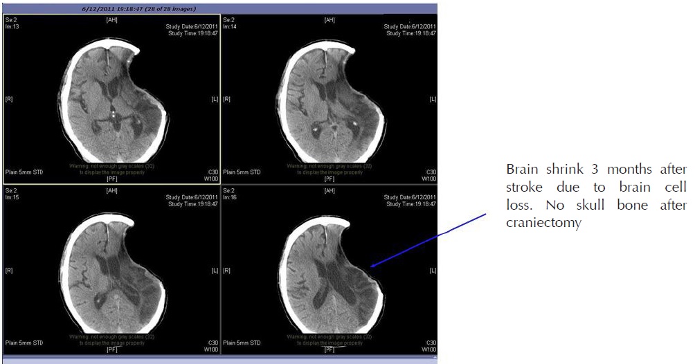
2. Cranial Decompressive Surgery: Craniectomy +/- Lobectomy
Large territory strokes can cause significant oedema of the brain with secondary brain injury in surrounding tissue. This phenomenon is mainly encountered in strokes of the middle cerebral artery territory, and is also called “malignant cerebral infaction” because it carries a dismal prognosis. Relief of the pressure may be attempted with medication, but some require decompressive craniectomy (temporary surgical removal of the skull) +/- lobectomy with excision of infarcted brain tissue. This confers a marked improvement in the risk of death, although most survivors are disabled [25].
|
References
1. World Health Organisation (1978). Cerebrovascular Disorders (Offset Publications). Geneva
2. Kidwell CS, Warach S (December 2003). “Acute ischemic cerebrovascular syndrome: diagnostic criteria”. Stroke 34 (12): 2995–8
3. Saver JL (2006). “Time is brain - quantified”. Stroke 37 (1): 263–6
4. The World health report 2004. Annex Table 2: Deaths by cause, sex and mortality stratum in WHO regions, estimates for 2002. Geneva
5. Murray CJ, Lopez AD (1997). “Mortality by cause for eight regions of the world: Global Burden of Disease Study”. Lancet 349 (9061): 1269–76.
6. Ellekjær, H; Holmen J, Indredavik B, Terent A (November 1, 1997). “Epidemiology of Stroke in Innherred, Norway, 1994 to 1996 : Incidence and 30-Day Case-Fatality Rate”. Stroke 28 (11): 2180–2184
| ©2017 Asia Medical Specialists Limited. All rights reserved. |
