Juvenile and Adolescent Idiopathic Scoliosis FAQs
Juvenile & Adolescent Idiopathic Scoliosis FAQs
written by Dr Bobby NG
Last update on: 16th Oct 2020
What is scoliosis?
Scoliosis is a form of twisting of the spine. The term scoliosis is from the Ancient Greek term for ‘bending’. The medical term scoliosis is used to describe sideways bending, although the deformity is usually twisting of the spine in three dimensions. The term kyphosis refers to forwards bending of the spine, giving a hunched appearance.
Are there different types of scoliosis?
Yes. The most common types occur in teenage and old age, but different types can occur in babies and young children, and other types at any age.
Here are the most common types, listed according to age:
Babies and young children:
- Congenital scoliosis – the child is born with an abnormal spine, and usually the scoliosis gets worse as the child grows.
Children and teens:
- Juvenile and Adolescent idiopathic scoliosis – this means ‘scoliosis occurring in children and teenagers of unknown cause’. It’s the most common cause in young people, and the subject of these Juvenile & Adolescent Idiopathic Scoliosis FAQs.
- Neuromuscular – there is a disease affecting the strength of the muscles, so they cannot support the spine eg muscular dystrophy.
- Syndromic scoliosis – scoliosis associated with specific diseases, e.g. Neurofibromatosis and Kabuki syndrome.
Young adults:
- Sciatic scoliosis – a slipped disc causes pain and associated twisting of the spine.
Elderly people:
- Degenerative scoliosis – the discs, and sometimes also the bones, collapse due to wear and osteoporosis, and the spine becomes twisted. This is the subject of the Degenerative Scoliosis FAQs.
Any age:
- Infection;
- Tumour;
- Leg Length Difference;
- Other causes.
How is scoliosis diagnosed?
Initially, simply by looking to see if the spine is straight.
Normally, the spine is almost perfectly straight, and we can see this when we look at a persons back. The trunk (or torso) is symmetrical and the shoulders are level in a normal person. If a scoliosis is present, the back will be curved.
If the curve is small the deformity may be mild and subtle and escapes detection, especially when clothed.
When the curve is moderate there may be some prominence of the rib cage on one side, called a rib hump in the chest region or a waist hump in the low back region.
If the curve is severe, there will be obvious deformity.
The rib or waist hump is usually more obvious if the patient bends forwards, and this can be measured with a scoliometer – a device similar to a spirit level.
An X-ray will demonstrate the problem. It should be a special X-ray of the whole spine, taken with the patient standing.
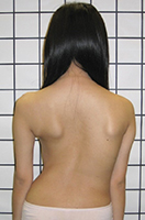
Figure 1a Right thoracic scoliosis showing right rib hump, asymmetry of the shoulders and shifting of the trunk resulting in a left ‘axillary gap’ between the arm and the body. |
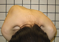
Figure1b Forward bending test showed prominent right rib hump resulting from the rotational deformity |
Are there different types of Juvenile & Adolescent Idiopathic Scoliosis?
The scoliosis can affect the whole spine from the neck down to the pelvis. One of the ways we describe scoliosis is based on the location and direction of the deformity. The location and direction helps to predict the prognosis.
What causes Juvenile & Adolescent Idiopathic Scoliosis?
We are not entirely sure, despite a vast amount of research!
We think it is a genetic condition with complex inheritance. The evidence for this includes that it affects identical twins with over 90% concordance although the severity and curve location may not be the same [1, 2].
Tell me more about the genetics?
Genome Wide Association Studies (GWAS) have identified many linkage chromosomes ( 6p, 6q, 8q,9q,10q,16q,17p,18q,19p,Xq) that harbour idiopathic scoliosis susceptibility [3]. More genes have also been associated with scoliosis such as the LBX1AS1, BCL-2, PAX/EPHA4, ASAP1 indicating multiple causes of idiopathic scoliosis [4, 5]. It seems this condition is genetically based due to mutation of gene units called Single Nucleotide Polymorphisms (SNPs) affecting the development of the spine. There is some tendency for these to occur in families.
How likely is my child to be affected with Juvenile or Adolescent Idiopathic Scoliosis?
Our large study of school screening in Hong Kong shows scoliosis affects about 3% of teenagers [6]. Of those who have scoliosis, about 10% would require brace treatment and 1% would end up requiring operation.
Scoliosis mainly affects girls. Mild scoliosis is about three times more common in girls than boys, and moderate to severe scoliosis is about eight times more common in girls than boys.
How do I know if my child has scoliosis?
Mild scoliosis escapes detection particularly when the child is wearing clothes. Parents may detect the abnormality when they have the opportunity to look at their child’s back, for example during summer when the child goes swimming.
The best way to know is to have an objective assessment by a medical professional or to participate in a school screening programme, which usually start at the age of 10. School screening may be especially valuable in Asia, where many teenagers dress quite conservatively, and scoliosis may not be noticed.
School screening is safe, simple, and not especially embarrassing. The child’s back can be observed through a special Moiré topography system, which highlights any asymmetry, and/or using a scoliometer as the child bends forward. It does not involve X-rays, though children are recommended to have X-rays if the screening test is positive.
How is scoliosis measured?
When scoliosis become significant, it will be visible on examination. The most objective method of assessment is to take an X-ray of the whole spine.
The scoliosis is described by its direction, the levels involved, the apex of the curve, and the size of the curve, which is known as the Cobb angle. For example the common location would be a right thoracic curve from T5 to T11 with apex located at T8,9 and Cobb angle of 25o.
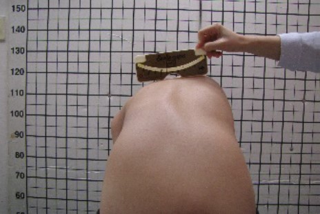
Idiopathic and other forms of ‘structural’ scoliosis have a special feature where the affected part of spine will twist toward the direction of deviation posteriorly and is called axial rotation. This will cause prominence of the rib cage, described as a ‘rib hump’. The severity of the rib hump corresponds to the degree of curvature of the scoliosis and is measured in the apical trunk rotation (ATR).
Figure 2 Measuring Apical Trunk Rotation with a scoliometer |
When does scoliosis become important?
Scoliosis becomes important when the Cobb angle gets to above 20o in a growing child. This is because with further growth the scoliosis is likely to progress. A study showed that with a curve greater than 20o in an immature child the risk of progression is 68%, while the risk of progression is only 22% if the child is more mature [7].
How do we estimate remaining growth, and thus the likelihood of scoliosis progression?
A number of factors are considered: age; skeletal maturity (based on X-rays); rate of growth; and, for girls, time since menarche (first menstrual period).
X-rays of the hand to look for the maturity of the growth plates in the hand and wrist, giving bone age, is a reliable method, but the conventional system describing the changes of these growth centres with age is complicated, requiring the use of reference charts. Our recent study using the thumb growth centres simplifies the assessment [8].
Risser’s sign is a method of assessment of the skeletal maturity of a person by looking at the maturity of the pelvic bone, which, conveniently, can usually be seen in the X-rays of the spine taken to assess the scoliosis [9]. The sequence of ossification follows a set pattern and this can be used to estimate the individual’s maturity and remaining growth potential. The Risser staging runs from I (immature) to V (fully skeletally mature).
Is scoliosis serious?
A very mild scoliosis does not matter at all, and the individual may not even be aware they have it.
A mild scoliosis may only be a minor cosmetic problem. They are not usually painful.
Unfortunately, severe scoliosis causes both cosmetic and functional problems.
Long-term studies show that with severe scoliosis, with a Cobb angle greater than 90o, leads to premature death from cardiopulmonary diseases [10].
Natural history studies of moderate (40-70 o) to severe (>70o) progressive scoliosis showed there was an increased death rate from age 40-80 years of age. This was particularly significant for the scoliosis of early onset [11].
When does scoliosis require treatment?
Treatment of scoliosis depends on the size of the curve and the age and maturity of the patient.
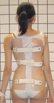
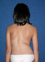
Brace treatment would be recommended for curves above 25o in a person with significant growth remaining. The brace would be worn 23 hours a day until the patient reached skeletal maturity, usually around 16 years of age.
Thereafter the risk of progression depends on the size of the curve. Larger curves, over 50o, are likely to continue to progress beyond skeletal maturity, whereas lesser curves usually do not.
For a curve that has progressed to more than 50o, operation is recommended.
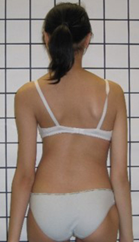
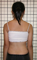
Figure 3 a & b, Girl with scoliosis treated with brace. |
What are the different kinds of operative treatment?
The current operative treatment of progressive scoliosis is to perform spinal fusion, straightening the spine, fusing (joining) the bones together and making the patient taller.
The exact technique depends on the type of scoliosis.
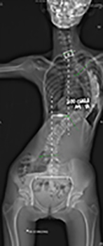
The most common type, thoracic scoliosis, is usually treated via a posterior approach – ie from behind. Current techniques involve placing multiple pedicle screws into each vertebral bone to achieve the best possible correction. Using modern navigation techniques improves the speed and accuracy of the procedures.
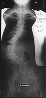
Fig 4A & B. X-rays showing thoracic scoliosis corrected by posterior spinal fusion with rods and pedicle screws. The screws were accurately placed into the spine using computer navigation. |
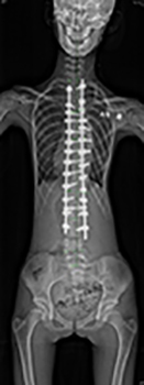
Spinal Fusion can also be done with anterior spinal fusion. This is mainly used for scoliosis that occurs in the mid spine region called the thoracolumbar scoliosis. This is very effective in correcting the three dimensional deformity of the spine and usually gives a near-perfect correction.
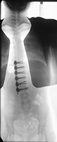
Figs 5A, B, C & D: Thoraco-Lumbar scoliosis corrected by anterior spinal fusion with vertebral body screws and dual rod system. |
What are the complications of operative treatment?
Operations on Idiopathic scolioisis are very safe, with total risk of significant complications around 2%. In a large survery of its members, the Scoliosis Research Society reported the risk of nerve injury at 0.75% .
What is the outcome of treatment of scoliosis?
The outcome of operative treatment has been comprehensively studied. In one study the quality of life of patients who had been treated by operation or brace was the same as normal controls [13].12 The improvement in lung function of patients treated by operation or brace was maintained up to 25 years after treatment [14].
Our recent studies of patients undergoing brace treatment [15] and also posterior spinal fusion surgery [16] showed excellent results for quality of life.
References
- Inoue, M., et al., Idiopathic scoliosis in twins studied by DNA fingerprinting: the incidence and type of scoliosis. J Bone Joint Surg Br, 1998. 80(2): p. 212-7.
- Weiss, H.R., Idiopathic scoliosis: how much of a genetic disorder? Report of five pairs of monozygotic twins. Dev Neurorehabil, 2007. 10(1): p. 67-73.
- Wise, C.A., et al., Understanding genetic factors in idiopathic scoliosis, a complex disease of childhood. Curr Genomics, 2008. 9(1): p. 51-9.
- Takahashi, Y., et al., A genome-wide association study identifies common variants near LBX1 associated with adolescent idiopathic scoliosis. Nat Genet, 2011. 43(12): p. 1237-40.
- Zhu, Z., et al., Genome-wide association study identifies new susceptibility loci for adolescent idiopathic scoliosis in Chinese girls. Nat Commun, 2015. 6: p. 8355.
- Luk, K.D., et al., Clinical effectiveness of school screening for adolescent idiopathic scoliosis: a large population-based retrospective cohort study. Spine (Phila Pa 1976), 2010. 35(17): p. 1607-14.
- Lonstein, J.E. and J.M. Carlson, The prediction of curve progression in untreated idiopathic scoliosis during growth. J Bone Joint Surg Am, 1984. 66(7): p. 1061-71.
- Hung, A.L., et al., Validation Study of the Thumb Ossification Composite Index (TOCI) in Idiopathic Scoliosis: A Stage-to-Stage Correlation with Classic Tanner-Whitehouse and Sanders Simplified Skeletal Maturity Systems. J Bone Joint Surg Am, 2018. 100(13): p. 88.
- Risser, J.C., The Iliac apophysis; an invaluable sign in the management of scoliosis. Clin Orthop, 1958. 11: p. 111-9.
- Nilsonne, U. and K.D. Lundgren, Long-term prognosis in idiopathic scoliosis. Acta Orthop Scand, 1968. 39(4): p. 456-65.
- Pehrsson, K., et al., Long-term follow-up of patients with untreated scoliosis. A study of mortality, causes of death, and symptoms. Spine (Phila Pa 1976), 1992. 17(9): p. 1091-6.
- MacEwen, G.D., W.P. Bunnell, and K. Sriram, Acute neurological complications in the treatment of scoliosis. A report of the Scoliosis Research Society. J Bone Joint Surg Am, 1975. 57(3): p. 404-8.
- Danielsson, A.J., et al., Health-related quality of life in patients with adolescent idiopathic scoliosis: a matched follow-up at least 20 years after treatment with brace or surgery. Eur Spine J, 2001. 10(4): p. 278-88.
- Pehrsson, K., A. Danielsson, and A. Nachemson, Pulmonary function in adolescent idiopathic scoliosis: a 25 year follow up after surgery or start of brace treatment. Thorax, 2001. 56(5): p. 388-93.
